DON'T LET THE AGE DEFINE YOUR STRENGTH: LIVE A HEALTHY FUTURE WITH IR TECHNOLOGY
Osteoporosis is a systemic skeletal disorder characterized by compromised bone strength, low bone mass, and disruption of bone architecture, predisposing to an increased risk of fracture. The World Health Organization (WHO) defines osteoporosis as having a bone mineral density (BMD) at the hip or lumbar spine greater than 2.5 standard deviations below the young normal adult reference population.
Vertebral fractures are the most common osteoporotic fracture in postmenopausal women. It is estimated that there are 550,000 to 700,000 osteoporotic vertebral compression fractures (VCFs) annually, which account for ~27% of all osteoporotic fractures in both men and women. The incidence is difficult to quantify accurately as only 23 to 33% of these fractures are clinically evident.
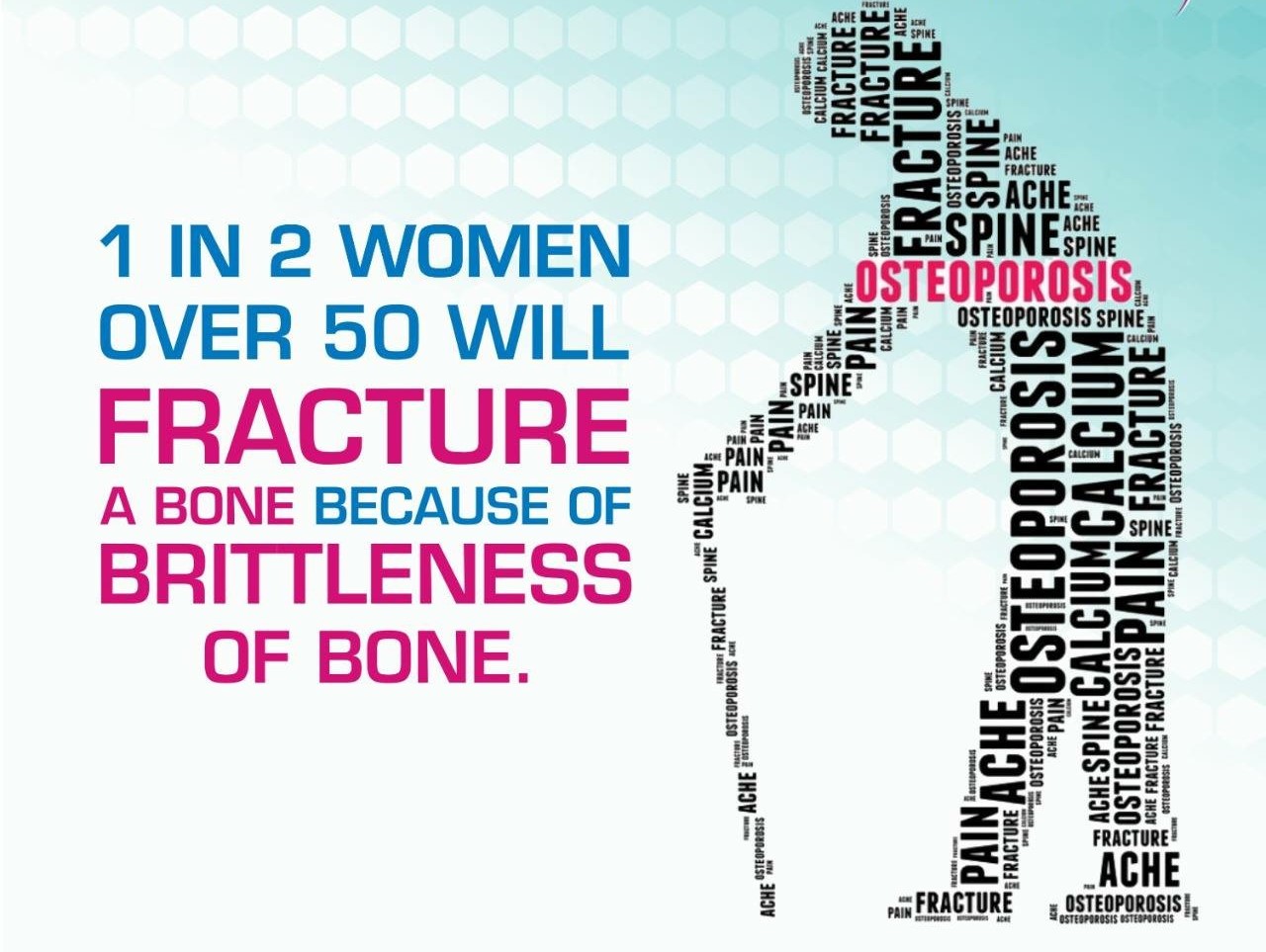
Osteoporosis occurs when the bones gradually become weak, increasing the risk of fractures. It is most common in women over the age of 60 and often leads to injuries in the hips, wrists, and spine. Causes can range from heredity, age, ethnicity, hormones (menopause, thyroid) and lifestyle choices.
The most common sites for osteoporotic fractures are in the back (vertebral compression fracture), the hip and the wrist.
The most common sites for osteoporotic fractures are in the back (vertebral compression fracture), the hip and the wrist.
Osteoporotic Spine Fractures Symptoms and Diagnosis
You may not recognize a fracture immediately. It’s important for those with chronic pain or patients who have been previously diagnosed with osteoporosis to be examined by a doctor. Symptoms include:
- Severe back pain
- The back pain worsened by standing or sitting for long periods of time
- Increasingly bad posture
- Initial diagnosis usually involves X-rays of the back. However; CT or MR scans may also be necessary.
TREATMENT: A HAPPY AND PAINFREE SOLUTION
THE INTERVENTIONAL RADIOLOGY WAY -


Interventional radiologists are board-certified physicians who deliver minimally invasive treatments with less risk, less pain and less recovery time than traditional surgery to treat conditions that impact a person’s quality of life, such as vertebral compression fractures.
- Vertebroplasty. Vertebroplasty is a minimally invasive treatment developed to treat pain caused by vertebral compression fractures, and has been safely performed since the late 1980s. Using X-ray imaging, an interventional radiologist inserts a needle into the collapsed vertebral body through a small incision in the skin. This image-guided technique allows the doctor to accurately access the fracture while minimizing trauma to surrounding tissue. A medical-grade liquid cement is then injected into the center of the vertebrae. As the cement solidifies, the broken bone is stabilized. The treatment is performed with the patient face-down and sedated for their comfort. Afterwards, many patients feel immediate relief from pain, and can be discharged home the same day.
- Kyphoplasty. Kyphoplasty is similar to vertebroplasty and is equally effective in stabilizing compression fractures. As with vertebroplasty, a needle is inserted into the fractured vertebra, using X-ray imaging. A balloon is then positioned into the collapsed bone and inflated to create a cavity for cement injection. The cement is injected into the cavity once the balloon is removed. Many patients feel immediate pain relief and are able to resume regular activities within a few days.


Comments
Post a Comment